

| Category | Pleural Empyema |
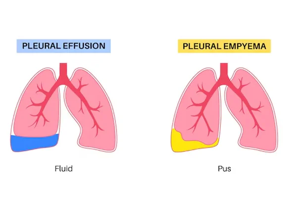
Pleural empyema is a serious condition in which infected fluid or pus collects in the pleural space, the area between the lungs and the chest wall. It commonly develops as a complication of pneumonia but can also occur after chest infections, surgery, trauma, or other conditions affecting the chest.
Timely diagnosis and treatment are important because an untreated empyema can become more difficult to manage and may affect normal lung expansion. If you are looking for pleural empyema treatment in Sarita Vihar, Dr. Saraansh Bansal provides specialist evaluation and personalised treatment guidance based on the severity and underlying cause of the condition.
The pleural space normally contains a very small amount of lubricating fluid that allows the lungs to move smoothly during breathing. When bacteria or other microorganisms infect this space, fluid can accumulate and become thick, infected, and pus-filled.
Pleural empyema may develop through different stages. Initially, the fluid may be relatively free-flowing. As the infection progresses, the fluid can become thicker and form separate pockets or collections. In more advanced cases, scar tissue may develop around the lung and restrict its expansion.
Because the condition can progress, appropriate medical assessment is important when empyema is suspected.
Pleural empyema most commonly develops following a bacterial infection in the lungs. Other possible causes and risk factors include:
The exact cause needs to be established because treatment may differ depending on the underlying infection.
Symptoms can develop gradually or become noticeable during an acute infection. Common symptoms include:
Some patients may continue to experience symptoms after treatment for pneumonia. Persistent fever, breathlessness, or chest pain following a respiratory infection should be evaluated.
Accurate diagnosis is an important part of pleural empyema treatment in Sarita Vihar. The doctor may begin by reviewing symptoms, medical history, previous infections, and treatment response.
Imaging tests such as a chest X-ray or CT scan can help identify fluid accumulation around the lung and provide information about its size and location. Ultrasound may also be useful for assessing pleural fluid and guiding drainage procedures.
In many cases, a sample of pleural fluid is collected for laboratory analysis. Testing the fluid can help identify infection and guide the choice of antibiotics.
Blood tests may also be performed to assess inflammation and infection.
Treatment usually focuses on controlling the infection and removing infected fluid from the pleural space. The appropriate treatment depends on the stage of the empyema, the patient's overall condition, and the characteristics of the fluid collection.
Antibiotics are an important part of treatment because empyema is generally associated with infection. The choice of antibiotic may depend on the suspected or identified organism, previous treatment, and laboratory results.
The duration of antibiotic treatment varies according to the individual clinical situation.
Infected pleural fluid often needs to be drained. A chest tube may be inserted into the pleural space to allow infected fluid to drain out.
Imaging guidance may be used to help position the drainage tube accurately, particularly when the collection is located in a difficult area.
In selected patients, medicines may be introduced into the pleural space to help break down thick collections and improve drainage. Whether this approach is appropriate depends on the characteristics of the empyema and the patient's condition.
If drainage and medical treatment do not adequately control the infection, surgery may be considered. Surgical options can include minimally invasive procedures such as video-assisted thoracoscopic surgery (VATS) or, in more advanced cases, other surgical approaches.
The goal is to remove infected material, release the lung when necessary, and help restore normal lung expansion.
Pleural empyema can become more organised over time. Thick infected fluid and scar tissue may make drainage more difficult and can restrict lung expansion.
Early assessment allows the medical team to determine the stage of the condition and select an appropriate treatment strategy. Patients recovering from pneumonia who develop persistent fever, chest pain, breathlessness, or worsening symptoms should seek medical evaluation.
Recovery depends on the severity of the infection, the amount of pleural fluid, the treatment required, and the patient's general health.
Patients may need follow-up imaging and clinical examinations to confirm that the infection is resolving and the lung is expanding appropriately. Antibiotics should be taken exactly as prescribed, and any drainage tube or wound requires appropriate care.
Adequate nutrition, hydration, rest, and gradual return to activity may support recovery. Breathing exercises or pulmonary rehabilitation may be recommended in selected cases.
Patients should contact their medical team if they develop worsening breathlessness, persistent fever, increasing chest pain, or other new symptoms.
Pleural empyema can require a combination of antibiotics, drainage, imaging, and sometimes surgical intervention. Dr. Saraansh Bansal focuses on detailed assessment and personalised treatment planning based on the severity and characteristics of each patient's condition.
For patients seeking pleural empyema treatment in Sarita Vihar, specialist consultation can help determine the cause of pleural infection, assess the extent of fluid collection, and identify appropriate treatment options.
Persistent symptoms after pneumonia or another chest infection should not be ignored. Pleural empyema can interfere with breathing and may require timely drainage and infection control.
Dr. Saraansh Bansal provides evaluation and treatment guidance for patients with pleural empyema in Sarita Vihar. The treatment approach is tailored according to the patient's symptoms, diagnostic findings, infection severity, and response to treatment.
If you have been diagnosed with pleural empyema or have persistent symptoms following a chest infection, a specialist consultation can help determine the next appropriate step.
Pleural empyema is an infection in the pleural space that causes infected fluid or pus to collect around the lung.
It most commonly develops as a complication of pneumonia or another chest infection. It can also occur after chest surgery, trauma, or certain other infections.
Some cases can be managed with antibiotics and drainage. However, surgery may be required when the infection does not respond adequately to these treatments or when thick, organised collections prevent effective drainage.
Diagnosis may involve chest X-ray, CT scan, ultrasound, blood tests, and analysis of pleural fluid obtained through drainage or aspiration.
Recovery varies depending on the severity of infection and the treatment required. Some patients recover relatively quickly, while complicated cases may require prolonged treatment and follow-up.
Persistent fever, chest pain, breathlessness, or worsening symptoms after pneumonia or another chest infection should be medically evaluated.
Dr. Saraansh Bansal provides specialist consultation and personalised treatment guidance for pleural empyema in Sarita Vihar.


